Showing posts with label scientists. Show all posts

Monday, 4 October 2010

Scientists achieve highest-resolution MRI of a magnet

COLUMBUS, Ohio -- In a development that holds potential for both data storage and biomedical imaging, Ohio State University researchers have used a new technique to obtain the highest-ever resolution MRI scan of the inside of a magnet.

Chris Hammel, Ohio Eminent Scholar in Experimental Physics, and his colleagues took a tiny magnetic disk -- measuring only 2 micrometers (millionths of a meter) across and 40 nanometers (billionths of a meter) thick – and were able to obtain magnetic resonance images its interior.

The resulting image -- with each "pixel" one tenth the size of the disk itself -- is the highest-resolution image ever taken of the magnetic fields and interactions inside of a magnet.
Highest-Resolution MRI Of A Magnet

Caption: Researchers at Ohio State University have developed a new type of magnetic resonance that can see inside magnetic materials. Here, slight variations in the structure of a thin magnetic film are evident as variations in ferromagnetic resonance frequency, represented by changes in color. Above the film is a representation of a polarized magnetic tip that scans the material.

Credit: Image courtesy of Ohio State University. Usage Restrictions: News Media use only.
Why look inside magnets? Because studying the material's behavior at these tiny scales is key to incorporating them into computer chips and other electronic devices.

The researchers report their findings in the August 12 issue of the journal Nature.

In 2008, Hammel's team debuted a new kind of high-resolution scanning system that combines three different kinds of technology: MRI, ferromagnetic resonance, and atomic force microscopy.

Ferromagnets -- the type of magnet used in this study -- are magnets made of ferrous metal such as iron. Common household refrigerator magnets are ferromagnets.

Because ferromagnets retain a particular polarization once magnetized, they are already essential components in today's computers and other electronics, where they provide data storage alongside computer chips.
But smaller magnets built directly into a computer chip could do even more, Hammel explained.

"We know that shrinking these magnets to the nanoscale and building them directly inside electronics would enable these devices to do more, and with less power consumption," Hammel said. "But a key barrier has always been the difficulty of imaging and characterizing nanomagnets."

Typical MRI machines work by inducing a magnetic field inside non-magnetic objects, such as the body. Since ferromagnets are already magnetic, conventional MRI can't see inside them.

The combination technique that the Ohio State researchers invented is called "scanned probe ferromagnetic resonance imaging," or scanned probe FMRI, and it involves detecting a magnetic signal using a tiny silicon bar with an even tinier magnetic probe on its tip.

In Nature, they report a successful demonstration of the technique, as they imaged the inside of the magnetic disk 0.2 micrometers (200 nanometers) at a time. They used a thin film of a commercially available nickel-iron magnetic alloy called Permalloy for the disk.

"In essence, we were able to conduct ferromagnetic resonance measurements on a small fraction of the disk, then move our probe over a little bit and do magnetic resonance there, and so on," explained Denis Pelekhov, director of the ENCOMM NanoSystems Laboratory at Ohio State. "Using these results, we could see how the magnetic properties vary inside the disk."

Experts suspect that computer chips equipped with tiny magnets might one day provide high-density data storage. Computers with magnets in their central processing units (CPUs) would never have to boot up. The entire computer would be contained inside the CPU, making such devices even smaller and less power-hungry as well.

Hammel believes that the technique could one day be useful tool in biomedical research labs. Researchers could use it to study tissue samples of the plaques that form in brain tissues and arteries, and perhaps develop better ways of detecting them in the body. Knowing how these plaques form could advance studies of many diseases, including Alzheimer's and atherosclerosis. ###

Hammel and Pelekhov's co-authors on the paper include Inhee Lee, Yuri Obukhov, Gang Xiang,, Adam Hauser, Fengyuan Yang, and Palash Banerjee, all of the Department of Physics at Ohio State.

This research was funded by the Department of Energy.

Contact: Chris Hammel Hammel.7@osu.edu 614-247-6928 Ohio State University Denis Pelekhov (614) 292-9125; Pelekhov.1@osu.edu Written by Pam Frost Gorder, (614) 292-9475; Gorder.1@osu.edu


The brain speaks Scientists decode words from brain signals

SALT LAKE CITY, – In an early step toward letting severely paralyzed people speak with their thoughts, University of Utah researchers translated brain signals into words using two grids of 16 microelectrodes implanted beneath the skull but atop the brain.

"We have been able to decode spoken words using only signals from the brain with a device that has promise for long-term use in paralyzed patients who cannot now speak," says Bradley Greger, an assistant professor of bioengineering.

Because the method needs much more improvement and involves placing electrodes on the brain, he expects it will be a few years before clinical trials on paralyzed people who cannot speak due to so-called "locked-in syndrome."

The Journal of Neural Engineering's September issue is publishing Greger's study showing the feasibility of translating brain signals into computer-spoken words.
16-Microelectrode Grid

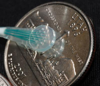
Caption: An array of 16 microelectrodes -- known as a microECoG grid -- is arranged in a four-by-four array and shown next to a US quarter-dollar coin with a Utah state design on its "tail" side. University of Utah researchers placed two such microelectrode grids over speech areas of a patient's brain and used them to decode brain signals into words. The technology someday might help severely paralyzed patients "speak" with their thoughts, which would be converted into a computerized voice.

Credit: Spencer Kellis, University of Utah. Usage Restrictions: None.

ECoG and microECoG Placement

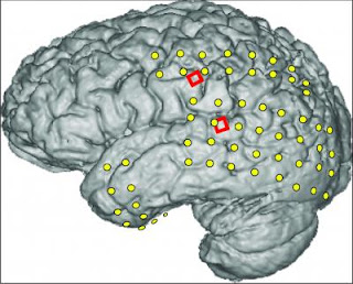
Caption: This magnetic resonance image (MRI) of an epileptic patient's brain is superimposed with the locations of two kinds of electrodes: conventional ECoG electrodes (yellow) to help locate the source of his seizures so surgeons could operate to prevent them, and two grids (red) of 16 experimental microECoG electrodes used to read speech signals from the brain. University of Utah scientists used the microelectrodes to translate brain signals into words -- a step toward devices that would let severely paralyzed people speak.

Credit: Kai Miller, University of Washington. Usage Restrictions: None.
The University of Utah research team placed grids of tiny microelectrodes over speech centers in the brain of a volunteer with severe epileptic seizures. The man already had a craniotomy – temporary partial skull removal – so doctors could place larger, conventional electrodes to locate the source of his seizures and surgically stop them.

Using the experimental microelectrodes, the scientists recorded brain signals as the patient repeatedly read each of 10 words that might be useful to a paralyzed person: yes, no, hot, cold, hungry, thirsty, hello, goodbye, more and less.

Later, they tried figuring out which brain signals represented each of the 10 words. When they compared any two brain signals – such as those generated when the man said the words "yes" and "no" – they were able to distinguish brain signals for each word 76 percent to 90 percent of the time.

When they examined all 10 brain signal patterns at once, they were able to pick out the correct word any one signal represented only 28 percent to 48 percent of the time – better than chance (which would have been 10 percent) but not good enough for a device to translate a paralyzed person's thoughts into words spoken by a computer.

"This is proof of concept," Greger says, "We've proven these signals can tell you what the person is saying well above chance. But we need to be able to do more words with more accuracy before it is something a patient really might find useful."

People who eventually could benefit from a wireless device that converts thoughts into computer-spoken spoken words include those paralyzed by stroke, Lou Gehrig's disease and trauma, Greger says. People who are now "locked in" often communicate with any movement they can make – blinking an eye or moving a hand slightly – to arduously pick letters or words from a list.

University of Utah colleagues who conducted the study with Greger included electrical engineers Spencer Kellis, a doctoral student, and Richard Brown, dean of the College of Engineering; and Paul House, an assistant professor of neurosurgery. Another coauthor was Kai Miller, a neuroscientist at the University of Washington in Seattle.
The research was funded by the National Institutes of Health, the Defense Advanced Research Projects Agency, the University of Utah Research Foundation and the National Science Foundation.

Nonpenetrating Microelectrodes Read Brain's Speech Signals

The study used a new kind of nonpenetrating microelectrode that sits on the brain without poking into it. These electrodes are known as microECoGs because they are a small version of the much larger electrodes used for electrocorticography, or ECoG, developed a half century ago.

For patients with severe epileptic seizures uncontrolled by medication, surgeons remove part of the skull and place a silicone mat containing ECoG electrodes over the brain for days to weeks while the cranium is held in place but not reattached. The button-sized ECoG electrodes don't penetrate the brain but detect abnormal electrical activity and allow surgeons to locate and remove a small portion of the brain causing the seizures.

Last year, Greger and colleagues published a study showing the much smaller microECoG electrodes could "read" brain signals controlling arm movements. One of the epileptic patients involved in that study also volunteered for the new study.

Because the microelectrodes do not penetrate brain matter, they are considered safe to place on speech areas of the brain – something that cannot be done with penetrating electrodes that have been used in experimental devices to help paralyzed people control a computer cursor or an artificial arm.

EEG electrodes used on the skull to record brain waves are too big and record too many brain signals to be used easily for decoding speech signals from paralyzed people.

Translating Nerve Signals into Words

In the new study, the microelectrodes were used to detect weak electrical signals from the brain generated by a few thousand neurons or nerve cells.

Each of two grids with 16 microECoGs spaced 1 millimeter (about one-25th of an inch) apart, was placed over one of two speech areas of the brain: First, the facial motor cortex, which controls movements of the mouth, lips, tongue and face – basically the muscles involved in speaking. Second, Wernicke's area, a little understood part of the human brain tied to language comprehension and understanding.

The study was conducted during one-hour sessions on four consecutive days. Researchers told the epilepsy patient to repeat one of the 10 words each time they pointed at the patient. Brain signals were recorded via the two grids of microelectrodes. Each of the 10 words was repeated from 31 to 96 times, depending on how tired the patient was. Then the researchers "looked for patterns in the brain signals that correspond to the different words" by analyzing changes in strength of different frequencies within each nerve signal, says Greger.

The researchers found that each spoken word produced varying brain signals, and thus the pattern of electrodes that most accurately identified each word varied from word to word. They say that supports the theory that closely spaced microelectrodes can capture signals from single, column-shaped processing units of neurons in the brain.

One unexpected finding: When the patient repeated words, the facial motor cortex was most active and Wernicke's area was less active. Yet Wernicke's area "lit up" when the patient was thanked by researchers after repeating words. It shows Wernicke's area is more involved in high-level understanding of language, while the facial motor cortex controls facial muscles that help produce sounds, Greger says.

The researchers were most accurate – 85 percent – in distinguishing brain signals for one word from those for another when they used signals recorded from the facial motor cortex. They were less accurate – 76 percent – when using signals from Wernicke's area. Combining data from both areas didn't improve accuracy, showing that brain signals from Wernicke's area don't add much to those from the facial motor cortex.

When the scientists selected the five microelectrodes on each 16-electrode grid that were most accurate in decoding brain signals from the facial motor cortex, their accuracy in distinguishing one of two words from the other rose to almost 90 percent.

In the more difficult test of distinguishing brain signals for one word from signals for the other nine words, the researchers initially were accurate 28 percent of the time – not good, but better than the 10 percent random chance of accuracy. However, when they focused on signals from the five most accurate electrodes, they identified the correct word almost half (48 percent) of the time.

"It doesn't mean the problem is completely solved and we can all go home," Greger says. "It means it works, and we now need to refine it so that people with locked-in syndrome could really communicate."

"The obvious next step – and this is what we are doing right now – is to do it with bigger microelectrode grids" with 121 micro electrodes in an 11-by-11 grid, he says. "We can make the grid bigger, have more electrodes and get a tremendous amount of data out of the brain, which probably means more words and better accuracy." ###

University of Utah Public Relations 201 Presidents Circle, Room 308 Salt Lake City, Utah 84112-9017 (801) 581-6773 fax: (801) 585-3350 www.unews.utah.edu

Contact: Lee J. Siegel leesiegel@ucomm.utah.edu 801-581-8993 University of Utah


(NanoTechnology)ORNL scientists help explain graphene mystery

OAK RIDGE, Tennessee, — Échelle du nanomètre simulations et recherches théoriques effectuée à Oak Ridge National Laboratory par le ministère de l'énergie apportent scientifiques plus proche pour réaliser le potentiel du graphene dans les applications électroniques.

Une équipe de recherche menée par de ORNL Bobby Sumpter, Vincent Meunier et Eduardo Cruz-Silva a découvert comment développent des boucles dans graphene, un matériau de faible poids haute résistance électrique conductif qui ressemble à un nid d'abeille à l'échelle atomique.

Les boucles structurelles qui font parfois au cours d'un processus de nettoyage graphene peuvent rendre le matériel inadapté pour les applications électroniques.Surmonter ces types de problèmes est d'un grand intérêt pour l'industrie électronique.

«Graphène est une étoile montante dans le monde de matériaux, compte tenu de son potentiel pour une utilisation dans des composants électroniques précis comme les transistors ou autres semi-conducteurs, a déclaré Bobby Sumpter, un scientifique de personnel au ORNL.
graphene layers

Les simulations ORNL démontrent comment les boucles (vu plus haut en bleu) entre les couches graphene peuvent être réduite à l'aide d'irradiation des électrons (en bas).
L'équipe dynamique moléculaire quantum permettant de simuler un graphene expérimentale, nettoyage de processus, comme expliqué dans un livre publié en lettres d'examen physique.Calculs effectués sur les superordinateurs ORNL a souligné les chercheurs à une étape intermédiaire négligée au cours du traitement.

Imagerie avec un microscope électronique de transmission, ou TEM, soumis le graphene à électrons irradiation, qui finalement a empêché la formation de boucle.Les simulations ORNL a montré qu'en injectant des électrons pour recueillir une image, les électrons changeaient simultanément structure du matériau.
«Prendre une photo avec un TEM n'est pas simplement une photo est prise,» a déclaré Sumpter.«Vous pourriez modifier l'image en même temps que vous cherchez à elle.»

La recherche s'appuie sur les conclusions présentées dans un livre de science 2009 (Jia et coll.), où Meunier et Sumpter aidé démontrer un processus qui nettoie les arêtes graphene en exécutant un courant à travers la matière dans un processus appelé Joule de chauffage.Graphène n'est aussi bon que l'uniformité ou la propreté de ses bords, qui déterminent comment efficacement le matériel peut transmettre des électrons. Meunier, a déclaré la possibilité de nettoyer efficacement les bords graphene est cruciale à l'utilisation de la matière en électronique.

«Imaginez vous disposez d'une voiture de sport fantaisie, mais vous vous rendez compte puis qu'il a roues carrés. Quelle bonne est-il ? C'est comme avoir des bords sur graphene, irréguliers "Meunier dit.

Études expérimentales récentes ont montré que le Joule chauffage processus peut conduire à des boucles indésirables qui se connectent graphene différents calques. Le papier PRL fournit une compréhension atomistique comment électrons irradiation d'un microscope électronique de transmission affecte le processus de nettoyage en empêchant la formation de la boucle de graphene.

«Nous pouvons nettoyer les bords et pas seulement que, nous sommes capables de comprendre pourquoi nous pouvons les, nettoyer» Meunier dit.

L'équipe de recherche inclus des scientifiques du Massachusetts Institute of Technology, Université Catholique de Louvain et Université Carlos III de Madrid.Sumpter et Meunier sont membres de l'informatique et en mathématiques division avec rendez-vous à l'Institut de la théorie des nanomatériaux au sein du Centre des sciences de matériaux Nanophase du ORNL.Cruz-Silva est un chercheur postdoctoral au ORNL.

Partie de ce travail a été appuyé par le Centre pour Nanophase Sciences des matériaux (CNMS) à ORNL.CNMS est l'un des cinq DOE échelle du nanomètre Science Research centres pris en charge par l'Office of Science DOE, installations de premier utilisateur national pour la recherche interdisciplinaire à l'échelle du nanomètre.Ensemble les NSRCs comprennent une suite d'installations complémentaires qui fournissent des chercheurs avec capacités de-la-pointe à fabriquer, processus, caractérisent et modéliser les matériaux de l'échelle du nanomètre et constituent le plus grand investissement dans l'infrastructure de l'initiative nationale de la nanotechnologie.Les NSRCs sont trouvent en Argonne du DOE, Brookhaven, Lawrence Berkeley, Oak Ridge et Sandia Los Alamos laboratoires nationaux.Pour plus d'informations sur les NSRCs DOE, visitez http://nano.energy.gov.ORNL est géré par UT-Battelle for Office de la science par le ministère de l'énergie.

Avec les médias : Morgan McCorkle Communications et relations de 865. 574. 7308