MiCRoSCoPE
من اساسيات تعليمنا وعملنا في قسم المختبرات الطبية هو استخدام المايكروسكوب بانواعه في تشخيص الحالات والكشف عن المايكروبات
ومن المعلوم ان للعين حد للرؤيا والوضح يقل مع صغر الجسم حتى تنعدم رؤية الاجسام الصغيرة جدا كما هو موضح بالصورة

ومن قديم الزمان وسالف العصر والاوان << تحسون انه بيقول قصة
لم يكن احد يعلم بوجود الاحياء الدقيقة حتى اكتشفت عن طريق ابو المايكرسكوب
Antony van Leeuwenhoek
الهولندي الجنسية

هو أول شخص لاحظ ووصف الكائنات المجهرية بدقة
عندما بدأ كتاجر اقمشة او صانع يجفف السلع وكان يستخدم عدسات تكبير لحساب الخيوط في القماش
علم نفسه صنع وصقل وتلميع عدسات صغيره جدا تصل الى قوة تكبير 270 X
وهي من اقوى عددسات التكبير ان ذاك
هذه العدسات ادت الى بناء مجاهر Leeuwenhoek التي اعتبرت اول المجاهر العلمية

Leeuwenhoek اول من اكتشف ووصف البكتيريا سنة 1674م
وقد رسمها كما في الشكل التالي

وهذا ماقاله لوين هوك في رسالة موجهة للعالم
My work, which I've done for a long time, was not pursued in order to gain the praise I now enjoy, but chiefly from a craving after knowledge, which I notice resides in me more than in most other men. And therewithal, whenever I found out anything remarkable, I have thought it my duty to put down my discovery on paper, so that all ingenious people might be informed thereof." - Anton Van Leeuwenhoek Letter of June 12, 1716
ولكن للاسف انك لاتجد أي شي من مجاهر لوين هوك فقد صنعت من الذهب والفضة وبيعت من قبل عائلته بعد وفاته
بعد ذلك العالم تبعه Robert Hooke العالم الانقليزي الذي طور المجهر
فلم يكن فقط مجرد مراقب بل طور المجهر فجعل له احتواء واحد ومصدر مستقل للضوء
وتوالت التطورات ف يصناعة المجهر بناء على التركيب البدائي الذي اسسه العالمان
والان دعونا ندرس المجهر بشكل علمي وتعاريف علمية
المحتوى الرئيسي للمايكروسكوب والتكبير هي العدسات
Lensesالعدسات
focus light rays at a specific place called the focal point
تعديل بؤرة اشعة الضوء في مكان معين يدعى النقطة المركزية
فإذا ماهي الـ focal point (الطول البؤري)؟
distance between center of lens and focal point is the focal length
هي المسافة بين مركز العدسة والنقطة المركزية
strength of lens related to focal length
short focal length Þmore magnification
لذا فقوة العدسة تتعلق بالطول البؤري
والتي تفسر بأن الطول البؤري القصير ينتج عنه تكبير اكثير

المجهر الضوئي وينقسم لعدة مجاهر :
The Light Microscope have many types:
bright-field microscope
dark-field microscope
phase-contrast microscope
fluorescence microscopes
are compound microscopes
image formed by action of ³2 lenses
The Bright-Field Microscope
المجهر الشائع الاستخدام
produces a dark image against a brighter background
تنتج صورة مظلمة وخلفية ساطعة
has several objective lenses
لها عدة عدسات موضعية
parfocal microscopes remain in focus when objectives are changed
total magnification
product of the magnifications of the ocular lens and the objective lens
عبارة عن نتيجة تكبير العدسة العينية والعدسة الموضعية


Microscope Resolution(الوضوح)
ability of a lens to separate or distinguish small objects that are close together
قدرة العدسة لتمييز او فصل الاجسام الصغيرة القريبة جدا
wavelength of light used is major factor in resolution
طول الموجة الضوئية المستعمل عامل رئيسي في الوضوح
shorter wavelength Þ greater resolution
طول الموجة الاقصر اكثر وضوح

working distance
distance between the front surface of lens and surface of cover glass or specimen
المسافة بين السطح الامامي للعدسة وسطح الغطاء الزجاجي او العينة

The Dark-Field Microscope
produces a bright image of the object against a dark background
صورة لامعه من الجسم ضد خلفية مظلمة
used to observe living, unstained preparations
يستخدم في التحضيرات الحية والغير مصبوغة
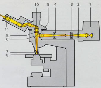
الصورة توضح التركيب الداخلي للمايكروسكوب


The Phase-Contrast Microscope
enhances the contrast between intracellular structures having slight differences in refractive index
يحسن المقارنة بين التركيبات الخلوية اتي لديها اختلاف بسيط في الـ refractive index
excellent way to observe living cells
الطريق الممتاز لملاحظة الخلايا الحية
الصورة توضح اتركيب الداخلي للمايكروسكوب


The Fluorescence Microscope
exposes specimen to ultraviolet, violet, or blue light
تعرض العينة للاشعة الفوق بنفسجية او البنفسجية او الزرقاء
specimens usually stained with fluorochromes
العينة تصبغ عادة بصبغة الـ fluorochromes
shows a bright image of the object resulting from the fluorescent light emitted by the specimen
تعطي صورة لامعة للجسم (مشعة) نتيجة للضوء المشع من العينة
الصورة توضح التركيب الداخلي للمايكروسكوب


ولاستخدام المجهر في التشخيص لابد من تحضير العينات قبل ذلك لاسباب عدة منها:
increases visibility of specimen
accentuates specific morphological features
preserves specimens
fixation
process by which internal and external structures are preserved and fixed in position
process by which organism is killed and firmly attached to microscope slide
heat fixing
preserves overall morphology but not internal structures
chemical fixing
protects fine cellular substructure and morphology of larger, more delicate organisms
Dyes and Simple Staining
dyes
make internal and external structures of cell more visible by increasing contrast with background
have two common features
chromophore groups
chemical groups with conjugated double bonds
give dye its color
ability to bind cells
simple staining
a single staining agent is used
basic dyes are frequently used
dyes with positive charges
e.g., crystal violet
Differential Staining
divides microorganisms into groups based on their staining properties
e.g., Gram stain
e.g., acid-fast stain
Gram staining
most widely used differential staining procedure
divides Bacteria into two groups based on differences in cell wall structure

Acid-fast staining
particularly useful for staining members of the genus Mycobacterium
e.g., Mycobacterium tuberculosis – causes tuberculosis
e.g., Mycobacterium leprae – causes leprosy
high lipid content in cell walls is responsible for their staining characteristics
Staining Specific Structures
Negative staining
often used to visualize capsules surrounding bacteria
capsules are colorless against a stained background
Spore staining
double staining technique
bacterial endospore is one color and vegetative cell is a different color
Flagella staining
mordant applied to increase thickness of flagella
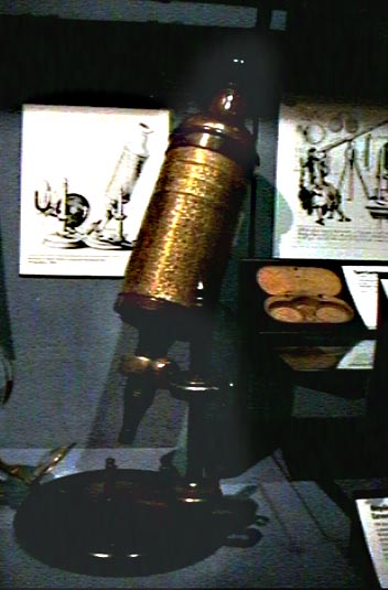
مجاهر اثرية


من اساسيات تعليمنا وعملنا في قسم المختبرات الطبية هو استخدام المايكروسكوب بانواعه في تشخيص الحالات والكشف عن المايكروبات
ومن المعلوم ان للعين حد للرؤيا والوضح يقل مع صغر الجسم حتى تنعدم رؤية الاجسام الصغيرة جدا كما هو موضح بالصورة
ومن قديم الزمان وسالف العصر والاوان << تحسون انه بيقول قصة
لم يكن احد يعلم بوجود الاحياء الدقيقة
حتى اكتشفت عن طريق ابو المايكرسكوبAntony van Leeuwenhoek
الهولندي الجنسية
هو أول شخص لاحظ ووصف الكائنات المجهرية بدقة
عندما بدأ كتاجر اقمشة او صانع يجفف السلع وكان يستخدم عدسات تكبير لحساب الخيوط في القماش
علم نفسه صنع وصقل وتلميع عدسات صغيره جدا تصل الى قوة تكبير 270 X
وهي من اقوى عددسات التكبير ان ذاك
هذه العدسات ادت الى بناء مجاهر Leeuwenhoek التي اعتبرت اول المجاهر العلمية
Leeuwenhoek اول من اكتشف ووصف البكتيريا سنة 1674م
وقد رسمها كما في الشكل التالي
وهذا ماقاله لوين هوك في رسالة موجهة للعالم
My work, which I've done for a long time, was not pursued in order to gain the praise I now enjoy, but chiefly from a craving after knowledge, which I notice resides in me more than in most other men. And therewithal, whenever I found out anything remarkable, I have thought it my duty to put down my discovery on paper, so that all ingenious people might be informed thereof." - Anton Van Leeuwenhoek Letter of June 12, 1716
ولكن للاسف انك لاتجد أي شي من مجاهر لوين هوك فقد صنعت من الذهب والفضة وبيعت من قبل عائلته بعد وفاته
بعد ذلك العالم تبعه Robert Hooke العالم الانقليزي الذي طور المجهر
فلم يكن فقط مجرد مراقب بل طور المجهر فجعل له احتواء واحد ومصدر مستقل للضوء
وتوالت التطورات ف يصناعة المجهر بناء على التركيب البدائي الذي اسسه العالمان
والان دعونا ندرس المجهر بشكل علمي وتعاريف علمية
المحتوى الرئيسي للمايكروسكوب والتكبير هي العدسات
Lensesالعدسات
focus light rays at a specific place called the focal point
تعديل بؤرة اشعة الضوء في مكان معين يدعى النقطة المركزية
فإذا ماهي الـ focal point (الطول البؤري)؟
distance between center of lens and focal point is the focal length
هي المسافة بين مركز العدسة والنقطة المركزية
strength of lens related to focal length
short focal length Þmore magnification
لذا فقوة العدسة تتعلق بالطول البؤري
والتي تفسر بأن الطول البؤري القصير ينتج عنه تكبير اكثير
المجهر الضوئي وينقسم لعدة مجاهر :
The Light Microscope have many types:
bright-field microscope
dark-field microscope
phase-contrast microscope
fluorescence microscopes
are compound microscopes
image formed by action of ³2 lenses
The Bright-Field Microscope
المجهر الشائع الاستخدام
produces a dark image against a brighter background
تنتج صورة مظلمة وخلفية ساطعة
has several objective lenses
لها عدة عدسات موضعية
parfocal microscopes remain in focus when objectives are changed
total magnification
product of the magnifications of the ocular lens and the objective lens
عبارة عن نتيجة تكبير العدسة العينية والعدسة الموضعية
Microscope Resolution(الوضوح)
ability of a lens to separate or distinguish small objects that are close together
قدرة العدسة لتمييز او فصل الاجسام الصغيرة القريبة جدا
wavelength of light used is major factor in resolution
طول الموجة الضوئية المستعمل عامل رئيسي في الوضوح
shorter wavelength Þ greater resolution
طول الموجة الاقصر اكثر وضوح
working distance
distance between the front surface of lens and surface of cover glass or specimen
المسافة بين السطح الامامي للعدسة وسطح الغطاء الزجاجي او العينة
The Dark-Field Microscope
produces a bright image of the object against a dark background
صورة لامعه من الجسم ضد خلفية مظلمة
used to observe living, unstained preparations
يستخدم في التحضيرات الحية والغير مصبوغة
الصورة توضح التركيب الداخلي للمايكروسكوب
The Phase-Contrast Microscope
enhances the contrast between intracellular structures having slight differences in refractive index
يحسن المقارنة بين التركيبات الخلوية اتي لديها اختلاف بسيط في الـ refractive index
excellent way to observe living cells
الطريق الممتاز لملاحظة الخلايا الحية
الصورة توضح اتركيب الداخلي للمايكروسكوب
The Fluorescence Microscope
exposes specimen to ultraviolet, violet, or blue light
تعرض العينة للاشعة الفوق بنفسجية او البنفسجية او الزرقاء
specimens usually stained with fluorochromes
العينة تصبغ عادة بصبغة الـ fluorochromes
shows a bright image of the object resulting from the fluorescent light emitted by the specimen
تعطي صورة لامعة للجسم (مشعة) نتيجة للضوء المشع من العينة
الصورة توضح التركيب الداخلي للمايكروسكوب
ولاستخدام المجهر في التشخيص لابد من تحضير العينات قبل ذلك لاسباب عدة منها:
increases visibility of specimen
accentuates specific morphological features
preserves specimens
fixation
process by which internal and external structures are preserved and fixed in position
process by which organism is killed and firmly attached to microscope slide
heat fixing
preserves overall morphology but not internal structures
chemical fixing
protects fine cellular substructure and morphology of larger, more delicate organisms
Dyes and Simple Staining
dyes
make internal and external structures of cell more visible by increasing contrast with background
have two common features
chromophore groups
chemical groups with conjugated double bonds
give dye its color
ability to bind cells
simple staining
a single staining agent is used
basic dyes are frequently used
dyes with positive charges
e.g., crystal violet
Differential Staining
divides microorganisms into groups based on their staining properties
e.g., Gram stain
e.g., acid-fast stain
Gram staining
most widely used differential staining procedure
divides Bacteria into two groups based on differences in cell wall structure
Acid-fast staining
particularly useful for staining members of the genus Mycobacterium
e.g., Mycobacterium tuberculosis – causes tuberculosis
e.g., Mycobacterium leprae – causes leprosy
high lipid content in cell walls is responsible for their staining characteristics
Staining Specific Structures
Negative staining
often used to visualize capsules surrounding bacteria
capsules are colorless against a stained background
Spore staining
double staining technique
bacterial endospore is one color and vegetative cell is a different color
Flagella staining
mordant applied to increase thickness of flagella
مجاهر اثرية
No comments:
Post a Comment